A - B - C - D - E - F - G - H - I - J - K - L - M - N - O - P - Q - R - S - T - U - V - W - X - Y - Z
N
Naturopath
Pronunciation: NAY-chur-uh-path
Definition: A naturopath is a health practitioner who applies the principles of Naturopathic Medicine, a distinct primary health care profession that emphasizes prevention, treatment, and optimal health through the use of therapeutic methods and substances that encourage individuals’ inherent self-healing process (vis medicatrix naturae). In a research context, Naturopathic Doctors (NDs) typically utilize a "Whole Systems" approach, integrating botanical medicine, clinical nutrition, and lifestyle modification to modulate neurobiology.
The Nootropic Research Interface
In the field of cognitive enhancement, naturopathic perspectives often drive the discovery and application of Phytoneurotropics. Their role in research is characterized by:
- Pleiotropic Pharmacology: Unlike "single-molecule" pharmaceutical research, naturopathic research often focuses on whole-plant extracts (e.g., Bacopa monnieri or Panax ginseng). These contain a complex matrix of alkaloids, saponins, and terpenes that may act synergistically across multiple pathways simultaneously—such as reducing neuroinflammation while enhancing cholinergic tone.
- The "Therapeutic Order": This is a clinical framework used to prioritize interventions. In nootropic research, a naturopathic approach ensures that foundational metabolic "leaks" (e.g., vitamin B¹² deficiency or circadian rhythm disruption) are addressed before introducing high-potency synthetic enhancers.
- Adaptogenic Theory: Naturopaths pioneered the study of Adaptogens—substances that increase a biological organism's resistance to stressors. Research in this area focuses on the modulation of the HPA (Hypothalamic-Pituitary-Adrenal) Axis to prevent the cognitive deficits associated with chronic cortisol elevation.
Professional Standards and Regulation
It is critical for researchers to distinguish between different levels of naturopathic training:
- Naturopathic Doctor (ND/NMD): A graduate of a four-year, accredited professional doctoral program. They are trained in the same basic sciences as MDs but specialize in natural therapeutics and integrative pharmacology.
- Traditional Naturopath: Typically refers to consultants who have not completed a formal, accredited medical program and whose scope of practice is generally limited to non-medical health counseling.
Primary Research Metrics
- Subjective Vitality Scales: Used to measure the "whole-person" impact of a nootropic protocol beyond simple memory scores.
- Allostatic Load: A composite measure of physiological "wear and tear" used to evaluate the long-term protective effects of naturopathic interventions.
- Synergy Quantitation: Using mathematical models to determine if the combination of compounds in a botanical extract provides a greater effect than the sum of its parts.
Research Note: When reviewing naturopathic literature, researchers must account for the "Entourage Effect." Because naturopaths often prefer crude or standardized extracts over isolated constituents, the pharmacokinetic profile may be more stable and less prone to the "crash" effects seen with isolated stimulants, albeit at the cost of more complex metabolic tracking.
NMDA Receptors (N-Methyl-D-Aspartate Receptor)
Pronunciation: en-em-dee-AY (EN-METH-ill-DEE-ah-SPAR-tayt) ree-SEP-tur
Definition: The NMDA receptor is an ionotropic glutamate receptor and a ligand-gated ion channel that is critical for the molecular mechanisms of learning and memory. It is unique among receptors because it is voltage-dependent and ligand-gated simultaneously. To open, the receptor requires both the binding of glutamate (and a co-agonist like glycine or D-serine) and a sufficient level of postsynaptic depolarization to expel a Magnesium (Mg²+) ion that normally blocks the pore. Once open, it allows for the influx of Calcium (Ca²+), which acts as a secondary messenger to trigger the signaling cascades required for Long-Term Potentiation (LTP).
The Nootropic Research Interface
In nootropic science, the NMDA receptor is the epicenter of "plasticity engineering." Research focuses on optimizing its function without crossing the threshold into excitotoxicity.
- NMDA Agonism & Co-agonism: Compounds that enhance NMDA function are studied for their ability to lower the "barrier to entry" for memory formation. Nootropics such as Sarcosine or D-Serine act on the glycine binding site to increase the probability of channel opening during learning events.
- The Magnesium Connection: Magnesium L-Threonate is a primary nootropic of interest because it increases the density of NMDA receptors in the hippocampus. By optimizing the "Magnesium block," it ensures that the receptors fire only in response to meaningful signals, thereby increasing the signal-to-noise ratio in the brain.
- Partial Agonism: Compounds like GLYX-13 (Rapastinel) are researched for their ability to act as "smart" modulators—activating the receptor enough to stimulate plasticity without causing the massive calcium influx that leads to cell death.
- Excitotoxicity & Protection: Chronic over-activation of NMDA receptors leads to an influx of Calcium so great that it triggers Apoptosis. Many neuroprotective nootropics (e.g., Memantine or Agmatine) act as uncompetitive antagonists, sitting in the channel to prevent it from staying open too long during pathological stress.
Molecular Subunits
NMDA receptors are heterotetramers, usually composed of two GluN1 subunits and two GluN2 (A, B, C, or D) subunits.
- GluN2B Subunit: Often called the "youth" subunit. It stays open longer and allows more calcium influx. Research into "upregulating" GluN2B expression is a major focus for reversing age-related memory decline.
- GluN2A Subunit: Becomes more prevalent as the brain matures. It has faster kinetics and is associated with the stabilization (pruning) of synapses.
Primary Research Metrics
- NMDA/AMPA Ratio: A measurement used in electrophysiology to determine the "plasticity potential" of a synapse. A higher ratio often indicates a synapse that is primed for strengthening.
- Excitotoxic Threshold: The concentration of glutamate required to induce 50% neuronal death; used to test the safety profile of NMDA-modulating nootropics.
- Receptor Density (Bmax): Quantified via radioligand binding or immunohistochemistry to determine if a nootropic has increased the total number of "learning gates" in a specific brain region.
Research Note: The NMDA receptor is a classic example of the Inverted-U dose-response. Too little activity results in an inability to form new memories (amnesia); too much activity results in neuronal "burnout" and death (excitotoxicity). The most effective nootropics in this category are those that stabilize the receptor in its "optimal" firing range.
Nerve Growth Factor
Pronunciation: NURV grohth FAK-tur
Definition: Nerve Growth Factor (NGF) is a neurotrophic factor and neuropeptide primary involved in the regulation of growth, maintenance, proliferation, and survival of specific target neurons. As the prototypic member of the Neurotrophin family, NGF acts by binding with high affinity to the Tropomyosin receptor kinase A (TrkA) and with low affinity to the p75 neurotrophin receptor (p75NTR). In the Central Nervous System (CNS), NGF is the primary trophic support for basal forebrain cholinergic neurons (BFCNs), which are the main source of acetylcholine for the hippocampus and neocortex.
The Nootropic Research Interface
In nootropic science, NGF is a central target for "bottom-up" cognitive repair and long-term neuroprotection. Because it cannot easily cross the blood-brain barrier (BBB) due to its high molar mass, research focuses on endogenous induction:
- Cholinergic Preservation: Since NGF is essential for the health of acetylcholine-producing neurons, inducing NGF is a primary strategy for treating Mild Cognitive Impairment (MCI). Without sufficient NGF, these neurons undergo atrophy, leading to the memory deficits characteristic of Alzheimer's Disease.
- Hericenone and Erinacine Induction: The most researched NGF-related nootropic is Lion's Mane (Hericium erinaceus). It contains low-molecular-weight compounds (erinacines and hericenones) that cross the BBB and stimulate the brain's natural production of NGF.
- Retrograde Signaling: NGF is produced by target tissues (like the hippocampus) and is captured by the axon terminals of projecting neurons. It is then transported "backward" (retrograde) to the cell body. Nootropics that optimize this Retrograde Transport are studied for their ability to maintain the "functional loop" between memory centers and executive centers.
The Dual-Receptor Mechanism
- TrkA Activation (Pro-Survival): Binding to TrkA triggers the MAPK/ERK and PI3K/Akt pathways, which promote neuronal survival, neurite outgrowth, and synaptic plasticity.
- p75NTR Activation (The "Pruning" Switch): In the absence of TrkA, or when binding to "pro-NGF" (the precursor molecule), p75NTR can initiate the JNK pathway, leading to programmed cell death. Research focuses on ensuring the Pro-NGF/Mature-NGF ratio favors the mature, survival-promoting form.
Primary Research Metrics
- Neurite Outgrowth Assay: A classic in vitro test where neurons are treated with a nootropic to see if they grow longer, more complex "branches" (dendrites and axons) via NGF induction.
- Choline Acetyltransferase (ChAT) Activity: An enzyme marker used to verify that NGF induction has successfully improved the functional capacity of cholinergic neurons.
- ELISA (Enzyme-Linked Immunosorbent Assay): Used to quantify the exact concentration of NGF in brain tissue or cerebrospinal fluid following administration of a secretagogue.
Research Note: While BDNF is often cited as the "fertilizer" for synapses and general plasticity, NGF is more specialized. It is the "specialized life support" for the cholinergic system. A nootropic stack that targets both (e.g., Lion's Mane for NGF and Bacopa for BDNF) is often considered a gold standard for comprehensive structural neuro-optimization.
Neurons
Pronunciation: NUR-onz
Definition: Neurons are the fundamental structural and functional units of the nervous system, specifically designed for the rapid transmission of information via electrical and chemical signals. They are post-mitotic, excitable cells characterized by a unique morphology—consisting of a soma (cell body), dendrites (input receivers), and a single axon (output transmitter). Unlike most other cells, neurons maintain a high electrochemical gradient across their membranes, which is discharged during an action potential to facilitate the release of neurotransmitters across the synaptic cleft.
The Nootropic Research Interface
In nootropic science, neurons are the primary "hardware" being optimized. Most cognitive enhancers function by modulating the environment, metabolism, or signaling efficiency of these cells:
- Bioenergetics & ATP: Neurons are metabolic "gas guzzlers," consuming a disproportionate amount of glucose and oxygen relative to their mass. Nootropics like Creatine or CoQ10 target the mitochondrial density within the neuron to prevent "metabolic failure" during high-intensity cognitive tasks.
- Neurogenesis & Survival: While most neurons are created during development, specific regions like the Hippocampus retain the ability to generate new neurons (Adult Neurogenesis). Nootropics that increase BDNF or NGF (e.g., Lion's Mane) are researched for their ability to promote the birth and integration of these new cells into existing circuits.
- Synaptic Plasticity: The "strength" of a neuron is measured by its ability to change its connections. Nootropics targeting LTP (Long-Term Potentiation) focus on the dendrites’ ability to sprout new spines, effectively increasing the neuron’s "surface area" for data reception.
- Membrane Fluidity: The neuronal membrane is composed of a phospholipid bilayer. Nootropics like Phosphatidylserine and Omega-3 fatty acids are used to maintain the "fluidity" of this membrane, ensuring that receptor proteins and ion channels can move and function with high efficiency.
Functional Classification in Research
- Excitatory Neurons: Primarily use Glutamate to increase the likelihood of the next neuron firing. Most "stimulatory" nootropics target these pathways.
- Inhibitory Neurons: Primarily use GABA to dampen neural activity, preventing excitotoxicity and "neural noise."
- Modulatory Neurons: Release "volume-control" chemicals like Dopamine, Acetylcholine, or Serotonin that alter the state of large neural networks simultaneously.
Primary Research Metrics
- Dendritic Spine Density: A measure of a neuron's connectivity; often used to quantify the "structural" benefits of a chronic nootropic protocol.
- Firing Rate (Hz): The frequency of action potentials; used to measure the acute "stimulatory" effect of a compound.
- Threshold Potential (mV): The voltage level required to trigger a neuron. Nootropics may "lower" this threshold to make a neuron more responsive or "raise" it to protect against over-excitation.
Research Note: It is a common misconception that "more neurons" equals "more intelligence." In nootropic research, the emphasis is shifted toward Connectivity and Synchronization. A brain with fewer, highly efficient, and well-myelinated neurons will consistently outperform a brain with more neurons that lack synchronized firing patterns.
Neurogenesis
Pronunciation: NUR-oh-JEN-uh-sis
Definition: Neurogenesis is the multi-stage biological process by which new functional neurons are generated from neural stem cells (NSCs) and progenitor cells. While once thought to occur only during embryonic development, it is now established that Adult Hippocampal Neurogenesis (AHN) persists throughout the human lifespan, primarily within the subgranular zone (SGZ) of the dentate gyrus and the subventricular zone (SVZ). The process encompasses cell proliferation, migration, differentiation, and the critical phase of synaptic integration into existing neural circuits.
The Nootropic Research Interface
In nootropic science, neurogenesis is the "long-game" of cognitive enhancement. Unlike stimulants that modulate existing neurotransmitters, pro-neurogenic compounds aim to increase the actual "computational density" of the brain.
- The Neurogenic Reserve: Research suggests that increasing the rate of AHN enhances "pattern separation"—the ability to distinguish between similar memories. Nootropics like Bacopa monnieri and Ginkgo biloba are studied for their ability to protect these nascent neurons during their vulnerable maturation phase.
- Trophic Factor Induction: Neurogenesis is heavily dependent on BDNF (Brain-Derived Neurotrophic Factor) and NGF. Nootropics that act as "mimetics" or secretagogues for these proteins (e.g., 7,8-DHF or Lion's Mane) are primary candidates for reversing the age-related decline in neurogenic rate.
- Environmental Enrichment & Allostasis: Chronic stress and high cortisol levels are potent inhibitors of neurogenesis. Nootropic adaptogens (e.g., Ashwagandha) are researched for their "permissive" effect—shielding the hippocampus from cortisol to allow baseline neurogenesis to resume.
- Stem Cell Niche Modulation: The "microenvironment" of the SGZ dictates whether a stem cell becomes a neuron or a glial cell. Research into Polyphenols (like Resveratrol or Curcumin) focuses on their ability to bias stem cell differentiation toward the neuronal lineage.
The Lifecycle of a New Neuron
- Proliferation: An NSC divides; one remains a stem cell, the other becomes a progenitor.
- Differentiation: The progenitor cell commits to becoming a neuron rather than a glial cell.
- Migration: The young "neuroblast" moves to its final position in the granular layer.
- Integration: The neuron extends dendrites and an axon, forming functional synapses. This stage is "use-it-or-lose-it"; without cognitive stimulation or nootropic support, the cell often undergoes apoptosis within weeks.
Primary Research Metrics
- BrdU Labeling: A synthetic nucleoside that integrates into the DNA of dividing cells, allowing researchers to "date" and count new neurons in histological samples.
- Doublecortin (DCX): A microtubule-associated protein expressed specifically in migrating neuronal precursors; used as a gold-standard biomarker for recent neurogenic activity.
- Volumetric MRI: In human subjects, an increase in the volume of the dentate gyrus is often used as a non-invasive proxy for successful neurogenic intervention.
Research Note: A common pitfall in nootropic marketing is the claim of "instant neurogenesis." In a research setting, the timeline for a new neuron to become a functional, signal-carrying member of a circuit is roughly 4 to 6 weeks. Therefore, pro-neurogenic nootropics require chronic administration and are often paired with "learning tasks" to ensure the survival of the new cells.
Neuroplasticity (Neural Plasticity)
Pronunciation: NUR-oh-plas-TISS-ih-tee
Definition: Neuroplasticity is the intrinsic ability of the brain to reorganize its structure, functions, and connections. It is an umbrella term encompassing several distinct biological processes: synaptic plasticity (changes in the strength of existing connections), structural plasticity (the physical growth or pruning of axons and dendrites), and functional plasticity (the cortical remapping of tasks from damaged areas to healthy ones). Unlike the legacy "static" view of the adult brain, neuroplasticity confirms that the neural "connectome" is in a state of constant flux, mediated by gene expression, de novo protein synthesis, and bioenergetic availability.
The Nootropic Research Interface
In the context of cognitive enhancement, neuroplasticity is the primary "mechanism of action" for long-term improvement. Nootropic interventions aim to increase the brain’s Plasticity Ceiling—the limit at which it can effectively adapt to new information.
- Metabolic Support for Rewiring: Structural changes (like forming new dendritic spines) are energy-intensive. Nootropics that enhance Mitochondrial function provide the $ATP$ necessary for the high-speed protein synthesis required during plastic windows.
- The "Permissive" State: Certain nootropics create a "permissive environment" for change. For example, Magnesium L-Threonate optimizes the density of NMDA Receptors, making the brain more sensitive to "coincidence detection" and lowering the threshold for Long-Term Potentiation (LTP).
- BDNF Modulation: Brain-Derived Neurotrophic Factor is the primary signal for plastic change. Nootropics like 7,8-DHF (a TrkB agonist) or Lion's Mane are studied for their ability to mimic or induce BDNF, effectively "softening" the neural circuitry to allow for easier reorganization.
- Epigenetic Regulation: Research into HDAC Inhibitors (such as certain dietary polyphenols) focuses on their ability to temporarily reopen "critical periods" of accelerated learning by altering chromatin accessibility.
Hierarchy of Plastic Change
- Chemical Plasticity: Rapid changes in neurotransmitter concentration and receptor sensitivity (milliseconds to seconds).
- Synaptic Plasticity: Strengthening or weakening of existing synapses via LTP or LTD (minutes to hours).
- Structural Plasticity: The physical growth of new synaptic "heads" (dendritic spines) or the pruning of unused ones (days to weeks).
- Functional Plasticity: Large-scale migration of cognitive functions to different cortical regions (months to years).
Primary Research Metrics
- VBM (Voxel-Based Morphometry): An MRI technique used to measure changes in gray matter volume as a proxy for structural plasticity in humans.
- fEPSP Slope: In electrophysiology, the slope of the field excitatory postsynaptic potential is the gold standard for measuring the "strength" of a potentiated synapse.
- Dendritic Arborization: The complexity of a neuron’s branching pattern, often quantified in animal models via Golgi staining or fluorescent labeling to assess the "growth" effects of a nootropic.
Research Note: While plasticity is generally a target for enhancement, "Maladaptive Plasticity" can lead to pathological states like chronic pain or tinnitus. Therefore, nootropic research often emphasizes "Directed Plasticity"—pairing a pharmacological enhancer with a specific learning stimulus to ensure the brain rewires itself toward a functional goal rather than "neural noise."
Neuroreceptor
Pronunciation: NUR-oh-ree-SEP-tur
Definition: A neuroreceptor is a specialized protein molecule situated on the cell membrane (postsynaptic or presynaptic) or within the cytoplasm of a neuron. It is designed to bind specifically to a ligand—such as a neurotransmitter, hormone, or exogenous nootropic compound. Upon binding, the receptor undergoes a conformational change that initiates a signal transduction cascade. Neuroreceptors are fundamentally categorized into two classes: Ionotropic (ligand-gated ion channels) which mediate rapid electrical shifts, and Metabotropic (G-protein coupled receptors) which trigger slower, long-lasting metabolic and genetic changes.
The Nootropic Research Interface
In neuropharmacology, the receptor is the primary site of "target engagement." Nootropic efficacy is defined by how a compound interacts with these proteins:
- Affinity vs. Efficacy: Research distinguishes between a nootropic's "grip" on the receptor (Affinity) and its ability to "flip the switch" (Efficacy). For instance, Racetams are often researched for their "positive allosteric modulation," which increases a receptor's sensitivity to its natural ligand without activating it directly.
- Up-regulation and Down-regulation: The brain maintains homeostasis by changing receptor density. Chronic use of certain enhancers (like stimulants) can lead to down-regulation (fewer receptors), resulting in tolerance. Conversely, "sensitizing" nootropics aim for up-regulation to restore cognitive baseline.
- Binding Site Specificity: Many receptors have multiple "pockets." Nootropics may bind to the orthosteric site (where the neurotransmitter goes) or an allosteric site (a secondary location). Allosteric modulators are highly prized in research for their "volume control" effect, which preserves the natural timing of neural firing.
Major Receptor Classes in Nootropics
Signal Transduction Pathways
- Ionotropic Effect: The receptor opens a pore, allowing ions (Ca²+, Na+, Cl-) to flow. This changes the neuron's electrical charge in milliseconds.
- Metabotropic Effect: The receptor activates a G-protein, which triggers "second messengers" like cAMP. This can lead to new protein synthesis and structural changes over minutes or hours.
Primary Research Metrics
- Ki Value: The inhibition constant; a lower Ki indicates a higher affinity for the receptor. Researchers use this to determine the "potency" of a new nootropic.
- Bmax: The maximum number of binding sites in a tissue sample; used to measure if a nootropic protocol has successfully increased receptor density.
- EC50: The concentration of a compound that induces a response halfway between the baseline and maximum; the standard measure of "functional potency."
Research Note: A common challenge in nootropic development is Receptor Cross-talk. A compound designed for a specific Serotonin receptor may inadvertently bind to others, leading to side effects. The goal of "high-selectivity" research is to hit the target receptor with surgical precision while ignoring the "off-target" proteins.
Neurofibrillary Tangles (NFTs)
Pronunciation: NUR-oh-FIB-ri-ler-ee TANG-gulz
Definition: Neurofibrillary Tangles are insoluble intracellular aggregates of hyperphosphorylated tau protein. In a healthy neuron, tau is a microtubule-associated protein (MAP) that stabilizes the "tracks" of the internal axonal transport system. In pathological states, tau undergoes chemical modification (excessive phosphorylation), causing it to detach from microtubules and misfold into paired helical filaments (PHFs). These filaments coalesce into "tangles" that physically obstruct the cytoplasm, leading to the collapse of the microtubule network, the cessation of nutrient transport, and eventual neuronal apoptosis.
The Nootropic Research Interface
In the study of "gero-nootropics" and neuroprotectants, NFTs represent a major obstacle to cognitive longevity. Research focuses on preventing the "tau-cascade" before tangles reach a critical mass:
- Tau Aggregation Inhibitors: Researchers are investigating compounds like Methylene Blue and its derivatives for their ability to dissolve PHFs or prevent their formation. These are studied for their potential to maintain "proteostasis"—the balance of protein production and clearance.
- Kinase Modulation: The hyperphosphorylation of tau is driven by enzymes like GSK-3β (Glycogen Synthase Kinase-3 Beta). Nootropics that inhibit overactive kinases, such as Lithium (micro-dose) or certain flavonoids, are researched for their ability to keep tau in its functional, non-tangled state.
- Autophagic Clearance: Once tangles form, the cell must clear them via autophagy (cellular recycling). Nootropics like Spermidine or Trehalose are being studied for their ability to "upregulate" the lysosomal machinery required to degrade these toxic protein aggregates.
- The "Spread" Mechanism: Emerging research suggests that pathological tau may spread from one neuron to another in a "prion-like" fashion. Nootropic strategies are being explored to stabilize the extracellular environment to prevent this trans-synaptic migration.
The Braak Staging Model
In clinical research, the progression of NFTs follows a predictable anatomical path known as Braak Stages:
- Stages I-II: Tangles are limited to the entorhinal cortex (early memory decline).
- Stages III-IV: Tangles spread to the hippocampus and limbic system (moderate cognitive impairment).
- Stages V-VI: Tangles reach the isocortex, leading to profound dementia and global cognitive failure.
Primary Research Metrics
- CSF Tau/p-Tau Levels: Measuring the concentration of total tau and phosphorylated tau in the cerebrospinal fluid; a high p-tau/Aβ42 ratio is a classic diagnostic signature.
- Tau-PET Imaging: Using radioligands (like Flortaucipir) to visualize the density and distribution of tangles in the living human brain.
- NFT Density: A histological count of tangles per square millimeter of brain tissue, used in post-mortem analysis to correlate "tangle burden" with the severity of cognitive symptoms.
Research Note: While Amyloid-Beta plaques are found outside the cell, Neurofibrillary Tangles are found inside the cell. Research indicates that while plaques may start the process, it is the density of tangles—the internal collapse of the neuron—that correlates most closely with the actual loss of memory and executive function.
Neurotransmitter
Pronunciation: NUR-oh-trans-MIT-er
Definition: A neurotransmitter is an endogenous chemical substance released by a neuron (the presynaptic cell) at a synapse that transmits a signal to an adjacent cell (the postsynaptic cell). To be classified as a neurotransmitter, a substance must meet the Daley Criteria: it must be synthesized in the neuron, present in the presynaptic terminal, released in amounts sufficient to exert a defined action, and possess a specific mechanism for removal/inactivation (such as enzymatic degradation or reuptake). These molecules function as the primary software of the brain, converting electrical action potentials into chemical signals that can bridge the synaptic cleft.
The Nootropic Research Interface
In nootropic science, neurotransmitters are the levers used to shift cognitive performance. Research focuses on four primary "interventions" to optimize neurochemistry:
- Precursor Loading: Providing the rate-limiting building blocks for synthesis. For example, L-Tyrosine is a precursor for Dopamine, and Alpha-GPC is a precursor for Acetylcholine. This is researched for its ability to prevent "transmitter depletion" during periods of high cognitive stress.
- Reuptake Inhibition: Extending the "residence time" of a transmitter in the synapse. Compounds like Polygala tenuifolia or certain synthetic stimulants are studied for their ability to block transporters (e.g., DAT, SERT), keeping the signal "active" for longer.
- Enzymatic Inhibition: Preventing the breakdown of the transmitter. A classic nootropic strategy involves using Acetylcholinesterase (AChE) inhibitors (like Huperzine A) to maintain high levels of Acetylcholine for improved memory encoding.
- Vesicular Release Modulation: Enhancing the "quantum" of transmitter released per nerve impulse. Nootropics in the Racetam family are researched for their ability to modulate ion channels and vesicle-docking proteins to ensure a robust "signal" is sent.
Functional Classification in Research
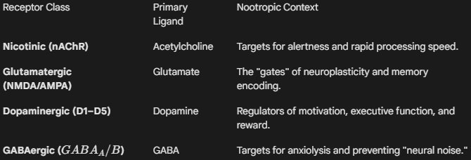
- Amino Acids: The "workhorses" of the brain. Glutamate (the primary excitatory signal) and GABA (the primary inhibitory signal) dictate the overall excitability of the cortex.
- Monoamines: The "state-shifters." Dopamine (motivation/focus), Serotonin (mood/executive control), and Norepinephrine (vigilance) modulate large-scale networks.
- Cholinergics: The "processors." Acetylcholine is the fundamental transmitter for synaptic plasticity, attention, and memory consolidation.
- Neuropeptides: The "fine-tuners." Larger molecules like Oxytocin or Enkephalins that modulate the sensitivity of the primary transmitter systems.
Primary Research Metrics
- Microdialysis: A technique used in vivo to measure the interstitial concentration of neurotransmitters in specific brain regions following nootropic administration.
- Turnover Rate: The speed at which a transmitter is synthesized and subsequently metabolized; a high turnover rate often indicates a system under high metabolic demand.
- Synaptic Clearance: The efficiency of transporters and enzymes in removing the transmitter from the cleft, a key factor in preventing "neural noise" or excitotoxicity.
Research Note: Many "crashes" associated with stimulants are the result of Neurotransmitter Exhaustion. A "rational" nootropic stack often pairs a stimulant (which increases release) with a precursor (which supports synthesis) and a neuroprotectant (which manages the metabolic cost) to ensure a sustainable increase in cognitive output.
Nootropic
Pronunciation: noh-oh-TROH-pik
Definition: A nootropic is a functional substance—whether synthetic, semi-synthetic, or biogenic—that enhances cognitive domains such as executive function, memory, creativity, or motivation in healthy individuals, while simultaneously exhibiting a neuroprotective profile. Unlike psychostimulants, which may improve performance at the cost of "metabolic debt" or systemic toxicity, a true nootropic must demonstrate a lack of significant side effects and low toxicity even at high dosages, specifically avoiding the induction of psychomotor agitation or sleep architecture disruption.
The Nootropic Research Interface
In modern neuropharmacology, the term "nootropic" describes a class of compounds that interface with the brain's homeostatic mechanisms to optimize the signal-to-noise ratio of neural transmission.
- The Giurgea Pillars: To be classified as a "classic" nootropic in research literature, a compound should ideally satisfy five criteria:
- Enhancement of learning and memory.
- Increased resistance of learned behaviors to conditions that tend to disrupt them (e.g., hypoxia, electroshock).
- Protection of the brain against physical or chemical injuries.
- Enhancement of the efficiency of tonic cortical/subcortical control mechanisms.
- Absence of typical pharmacological effects of neuropsychotropic drugs (e.g., sedation, motor stimulation).
- Bioenergetic Modulation: Research focuses on how nootropics increase cerebral metabolic rate without increasing oxidative stress. This often involves the upregulation of Mitochondrial efficiency or the enhancement of glucose utilization in the cerebral cortex.
- Integrative Connectivity: Unlike targeted "drugs" that might hit a single receptor, nootropics are often pleiotropic. They may simultaneously modulate Neurotransmitters (like Acetylcholine), increase Cerebral Blood Flow (CBF), and stimulate the release of neurotrophic factors like BDNF.
Categorization in Research
- Racetams: The foundational class (e.g., Piracetam, Aniracetam) known for modulating glutamate and acetylcholine receptors.
- Cholinesterase Inhibitors: Agents that prevent the breakdown of acetylcholine (e.g., Huperzine A).
- Adaptogens: Botanicals that modulate the HPA axis to maintain cognitive performance under stress (e.g., Rhodiola Rosea, Ashwagandha).
- Metabolic Enhancers: Compounds that improve the raw materials for brain function (e.g., Creatine, CoQ10).
Primary Research Metrics
- Pharmacokinetic Selectivity: The degree to which a substance targets specific cognitive pathways without systemic "off-target" effects.
- Psychometric Testing: Use of standardized tools like the Cambridge Neuropsychological Test Automated Battery (CANTAB) to quantify improvements in memory and attention.
- Neuroprotection Index: A measure of a compound's ability to preserve neuronal viability against stressors (ROS, excitotoxicity, or hypoxia) in in vitro models.
Research Note: A critical distinction in contemporary literature is the difference between a Nootropic and a Cognitive Enhancer. All nootropics are cognitive enhancers, but not all cognitive enhancers (like Amphetamines) are nootropics, as they lack the requisite neuroprotective safety profile and "non-stimulatory" nature.
Nootropic Stack
Pronunciation: noh-oh-TROH-pik STAK
Definition: A nootropic stack is the deliberate co-administration of two or more cognitive-enhancing substances to produce a synergistic or additive effect that exceeds the efficacy of any single constituent used in isolation. In a research context, a stack is a complex multi-component intervention designed to modulate distinct but interrelated neurobiological systems—such as pairing a cholinergic agonist with a high-affinity choline precursor to optimize the Acetylcholine lifecycle.
The Nootropic Research Interface
In clinical and self-directed research, the "stack" is the primary unit of optimization. Designing an effective stack requires an understanding of Pharmacodynamics (what the drugs do to the body) to prevent receptor competition or antagonistic interactions.
- Synergistic Stacking: Combining compounds that work through different mechanisms to achieve a single goal. A classic example is the L-Theanine and Caffeine stack; Caffeine increases alertness via adenosine antagonism, while L-Theanine modulates GABA and alpha-wave activity to mitigate the jitteriness and vasoconstriction associated with caffeine.
- Bioenergetic "Full-Cycle" Stacking: This strategy addresses both the trigger and the fuel. For instance, if a researcher uses a Racetam to increase glutamate receptor sensitivity (the trigger), they may "stack" it with Creatine or CoQ10 to ensure the mitochondria can meet the resulting increase in ATP demand (the fuel).
- The "Base" and "Finisher" Model: Most research-grade stacks utilize a "Base"—a foundational nutrient like an Omega-3 fatty acid or a B-Vitamin complex—paired with a "Finisher" or "Active Agent"—a high-potency compound like Noopept or Modafinil. This ensures that the brain has the structural raw materials necessary to support the accelerated signaling.
The Logic of the Stack
Researchers evaluate stacks based on three primary interactions:
- Additive (1 + 1 = 2): The combined effect is the sum of the individual parts.
- Synergistic (1 + 1 = 3): The combination produces a magnified response greater than their sum (e.g., Curcumin stacked with Piperine to increase bioavailability).
- Antagonistic (1 + 1 = 0.5): One compound inhibits the other, often due to competing for the same metabolic enzymes (e.g., CYP450) or the same receptor sites.
Primary Research Metrics
- Interaction Analysis: Using isobolograms to graphically determine if a combination of two nootropics is truly synergistic or merely additive.
- The "Washout" Period: The time required between changing stacks to ensure that the metabolites of a previous "Base" do not confound the data of a new intervention.
- Stack Efficiency Ratio (SER): A theoretical metric comparing the cognitive gain per milligram of a stack versus the constituent components.
Research Note: The primary risk in "untested" stacking is Metabolic Overload. When multiple compounds are metabolized by the same pathway (such as the Cytochrome P450 system in the liver), the "stack" can lead to unintended increases in blood-plasma concentrations, potentially turning a safe nootropic profile into a toxic one.
Noradrenaline (Norepinephrine) (NE)
Pronunciation: nor-uh-DREN-uh-lin
Definition: Noradrenaline is a catecholamine that functions as both a hormone and a neurotransmitter. In the Central Nervous System (CNS), it is synthesized primarily within the locus coeruleus (LC), a nucleus in the pons that sends extensive projections to the cerebral cortex, hippocampus, and amygdala. Synthesized from L-Tyrosine via the Dopamine precursor, noradrenaline binds to three main classes of adrenergic receptors: α1, α2, and β. Its primary function is to regulate "state-dependent" brain activity, shifting the neural landscape from a state of rest to one of high-vigilance, focused attention, and executive mobilization.
[Image showing the Locus Coeruleus and its widespread noradrenergic projections throughout the human brain]
The Nootropic Research Interface
In nootropic science, noradrenaline is the "gain control" of the brain. Research focuses on optimizing its levels to find the "Goldilocks zone" for cognitive throughput:
- Signal-to-Noise Ratio (SNR): Noradrenaline increases the "gain" of neurons. By activating postsynaptic α2-adrenoceptors in the prefrontal cortex, it strengthens relevant neural signals while simultaneously dampening background "noise." Nootropics that gently boost noradrenaline (e.g., L-Tyrosine or Mucuna Pruriens) are researched for their ability to maintain focus during sleep deprivation or high-stress tasks.
- The Yerkes-Dodson Inverted-U: Cognitive performance is dependent on noradrenergic tone. Too little noradrenaline results in drowsiness and poor integration; too much leads to anxiety and "cognitive "flexibility" loss. Many nootropics act as Adrenergic Modulators, attempting to shift the user toward the peak of the curve.
- Memory Consolidation: Noradrenaline release during an event signals to the hippocampus that the information is "important," triggering the molecular machinery of Long-Term Potentiation (LTP). This makes it a target for enhancing long-term retention.
- Vigilance and Arousal: Unlike "stimulants" that primarily hit dopamine (motivation), noradrenergic agents (e.g., Modafinil or Selective Norepinephrine Reuptake Inhibitors) are researched for their ability to sustain tonic arousal—the ability to stay awake and alert over long durations.
Molecular Biosynthesis
- L-Tyrosine → L-DOPA (via Tyrosine Hydroxylase)
- L-DOPA → Dopamine (via DOPA Decarboxylase)
- Dopamine → Noradrenaline (via Dopamine $\beta$-Hydroxylase)
Primary Research Metrics
- Tonic vs. Phasic Firing: Researchers distinguish between the constant "background" leak of noradrenaline (tonic) and the rapid bursts (phasic) associated with new stimuli. A high tonic/phasic ratio is often a marker of ADHD or distractibility.
- MHPG (3-methoxy-4-hydroxyphenylglycol): The primary metabolite of noradrenaline; measured in blood or urine as a proxy for central noradrenergic turnover.
- Pupillometry: Because the pupil's diameter is tightly linked to locus coeruleus activity, it is used in nootropic trials as a non-invasive, real-time metric of noradrenergic arousal.
Research Note: Many "energy" supplements prioritize noradrenaline release, but this can lead to "Peripheral Side Effects" (tachycardia, hypertension) because noradrenaline also acts in the sympathetic nervous system. Modern "smart" nootropics aim for central selectivity, attempting to boost brain-focus without over-stimulating the heart.