A - B - C - D - E - F - G - H - I - J - K - L - M - N - O - P - Q - R - S - T - U - V - W - X - Y - Z
G
Glial Cells
Pronunciation: GLEE-ul sel
Definition: Glial cells are the non-neuronal cellular components of the Central Nervous System (CNS) and Peripheral Nervous System (PNS) that do not produce electrical impulses but maintain homeostasis, provide myelination, and participate in signal transduction. Once thought to be passive support structures, glia are now recognized as integral partners in the "Tripartite Synapse," actively modulating the strength and longevity of neuronal connections.
The Nootropic Research Interface
In cognitive enhancement research, "Gliotropic" interventions are a growing frontier. Enhancing glial function is often a prerequisite for maximizing neuronal performance:
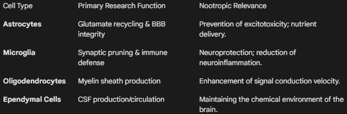
- Astrocyte-Mediated Metabolic Support: Astrocytes provide the primary energy source for neurons via the Astrocyte-Neuron Lactate Shuttle. Nootropics that optimize astrocytic glycogen storage or lactate transport (such as CoQ10 or certain Polyphenols) ensure that neurons have the "fuel" necessary for high-frequency firing.
- Glymphatic Clearance: During sleep, astrocytes shrink to facilitate the flow of cerebrospinal fluid through the brain, flushing out metabolic waste (like Amyloid-beta). Researchers study "recovery" nootropics (e.g., Melatonin, Magnesium L-Threonate) for their ability to enhance this glial-driven detoxification process.
- Microglial Modulation: Microglia act as the brain's resident immune cells. Chronic "low-grade" neuroinflammation—where microglia remain in a pro-inflammatory (M¹) state—is a primary cause of "brain fog" and cognitive decline. Nootropic adaptogens (like Curcumin or Longvida) are researched for their ability to shift microglia toward a neuroprotective (M²) anti-inflammatory phenotype.
- Oligodendrocyte Myelination: These cells wrap axons in myelin, which determines Processing Speed. Research into "remyelinating" agents focuses on improving the "hardware" speed of the CNS.
Functional Classification in the CNS
Mechanisms of Action
- Glutamate Clearance: Glia express transporters (like EAAT1/2) that vacuum up excess glutamate from the synapse. Without efficient glial function, neurons suffer from excitotoxic stress, which kills "focus" and leads to long-term damage.
- Synaptic Pruning: Microglia "eat" weak or redundant synapses. Nootropics that support healthy pruning ensure the brain remains efficient and isn't bogged down by "noisy" or inefficient neural pathways.
- Trophic Factor Secretion: Glial cells are a major source of BDNF (Brain-Derived Neurotrophic Factor) and GDNF (Glial-Derived Neurotrophic Factor), which are essential for the survival and growth of new neurons.
Primary Research Metrics
- GFAP (Glial Fibrillary Acidic Protein): A protein marker used to identify astrocytic activation or "gliosis" (scarring) in response to injury.
- Iba1 (Ionized calcium-binding adapter molecule 1): A marker used to visualize microglial morphology and determine if they are in an "active" (inflammatory) or "resting" state.
- D-Serine Levels: An amino acid co-agonist of the NMDA receptor primarily released by glia; used as a proxy for glial involvement in learning.
Research Note: When a subject experiences "diminishing returns" from a stimulant, it is often not the neurons that are exhausted, but the astrocytes that have run out of lactate or failed to clear the accumulating metabolic "exhaust" from the synaptic cleft.
Glutamate
Pronunciation: GLOO-tuh-mate
Definition: Glutamate (C⁵H⁹NO⁴) is an anionic form of the amino acid glutamic acid and serves as the most abundant excitatory neurotransmitter in the vertebrate nervous system. It is estimated that over 90% of the excitatory synapses in the human brain are glutamatergic. As the primary mediator of "fast" excitatory transmission, glutamate is essential for nearly all aspects of brain function, including sensory processing, motor control, and—crucially for nootropic research—the cellular mechanisms of learning and memory.
The Nootropic Research Interface
In the context of cognitive enhancement, glutamate is the "working fluid" of the brain’s computational engine. Research typically focuses on the modulation of its two primary ionotropic receptor classes:
- AMPA Receptors: Responsible for fast, "moment-to-moment" synaptic transmission. Nootropics known as AMPAkines (e.g., Aniracetam, Sunifiram) are researched for their ability to positively modulate these receptors, increasing the "clarity" of the signal and improving processing speed.
- NMDA Receptors: These act as "coincidence detectors," only opening when the neuron is sufficiently depolarized and glutamate is present. This is the gateway to Long-Term Potentiation (LTP). Many nootropic protocols aim to optimize NMDA receptor sensitivity to facilitate the "locking in" of new memories.
- The Glutamate-GABA Balance: Since glutamate is the precursor to GABA (the primary inhibitory neurotransmitter), research often focuses on the "Redox Status" and enzymatic activity (GAD) that converts one to the other to prevent over-excitation.
The Glutamate-Glutamine Cycle
Glutamate levels must be tightly regulated to prevent cellular damage. This is managed via a partnership between neurons and astrocytes (glial cells):
- Release: Glutamate is released into the synaptic cleft.
- Uptake: Excess glutamate is rapidly vacuumed up by astrocytes via EAAT (Excitatory Amino Acid Transporters).
- Conversion: Inside the astrocyte, glutamate is converted into the "inert" Glutamine.
- Recycling: Glutamine is sent back to the neuron, where it is converted back into active Glutamate for the next signal.
Primary Research Metrics
- E/I Ratio: The ratio of Excitation (Glutamate) to Inhibition (GABA). A "skewed" E/I ratio is a primary biomarker for brain fog, anxiety, or ADHD.
- MRS (Magnetic Resonance Spectroscopy): A non-invasive imaging technique used to measure the concentration of glutamate in specific brain regions in vivo.
- Glutamate Clearance Rate: A measure of how quickly the brain can "clean up" after a signal, often used to assess the health of the glial support system.
Risk Factor: Excitotoxicity
Because glutamate is so powerful, "more" is not always better. Excitotoxicity occurs when excessive glutamate over-stimulates NMDA receptors, leading to a massive influx of calcium (Ca²+) that can trigger neuronal apoptosis (cell death).
Research Note: Many "neuroprotective" nootropics (like Magnesium or Memantine) function by acting as "voltage-dependent blocks" on the NMDA receptor, preventing background "noise" from triggering excitotoxic pathways while still allowing for strong "learning" signals to pass through.
Gray Matter
Pronunciation: GR-ay MAT-ur
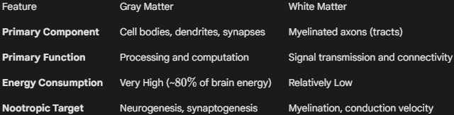
Definition: A major component of the Central Nervous System (CNS), consisting primarily of neuronal cell bodies (soma), unmyelinated axons, dendrites, glial cells (astrocytes and microglia), and capillaries. In the brain, gray matter is concentrated in the outer layer (cerebral cortex) and deep subcortical nuclei (such as the thalamus, basal ganglia, and hippocampus). Unlike white matter, which facilitates communication between regions, gray matter is the site of actual information processing, sensory perception, and executive decision-making.
The Nootropic Research Interface
In the study of cognitive enhancers, gray matter serves as a structural biomarker for "Cognitive Reserve." Researchers often track changes in Gray Matter Volume (GMV) to determine if a nootropic protocol is inducing genuine structural changes rather than just transient neurochemical shifts.
- Synaptogenesis and Dendritic Complexity: An increase in gray matter density is often interpreted as evidence of increased synaptic connections and dendritic branching. Nootropics that upregulate BDNF (Brain-Derived Neurotrophic Factor) or NGF (Nerve Growth Factor) are frequently evaluated by their ability to prevent or reverse the gray matter atrophy associated with aging or chronic stress.
- Cortical Thickness: This specific metric of gray matter is highly correlated with IQ and executive function. Research into "long-term" nootropics (e.g., Omega-3s, Bacopa monnieri, or intensive meditation) focuses on their capacity to maintain or increase cortical thickness in the Prefrontal Cortex.
- Metabolic Demand: Gray matter is the most metabolically expensive tissue in the body, consuming significantly more oxygen and glucose than white matter. Nootropics that enhance Cerebral Blood Flow or Mitochondrial Efficiency are essential for sustaining the high energy requirements of gray matter during intense "Deep Work" sessions.
Composition Comparison
Primary Research Metrics
- VBM (Voxel-Based Morphometry): A neuroimaging technique used to investigate focal differences in brain anatomy, specifically measuring the concentration or volume of gray matter in a 3D space.
- Cortical Mapping: Used to measure the depth and surface area of the gray matter "ribbon" across the cerebral hemispheres.
- Neuropil Density: A microscopic measurement of the space between cell bodies (filled with dendrites and synapses), which contributes to the overall volume of gray matter.
Research Note: While "more" gray matter is generally associated with higher cognitive function, the "Synaptic Pruning" phase in adolescence shows that a reduction in gray matter volume can also represent a maturation process where the brain becomes more efficient by removing redundant or "noisy" connections. In adult nootropic research, the goal is typically preservation of volume and optimization of synaptic density within that volume.